
Abdominal ultrasound
zł 270.00
Modern medicine utilizes numerous methods for internal body evaluation, among which ultrasound stands out for its maximum comfort and lack of discomfort. When planning a clinic visit, it is helpful to understand what ultrasound is and how the examination is performed.
Essentially, it is a safe imaging technology that assesses internal body structures using very high-frequency sound waves inaudible to the human ear. A dedicated device directs these imperceptible sound pulses into the body, which bounce off internal tissues as echo signals, and computer software converts the received data into a real-time, dynamic image on the screen within fractions of a second.
Our medical center provides ultrasound examinations of all organs for both adults and children. We help detect hidden health conditions at the earliest stages, before noticeable symptoms develop.
At „Lekarze w Warszawie”, the following types of ultrasound are available:
We use expert-grade digital systems that deliver high-resolution imaging. This allows our physicians to identify even microscopic deviations from the norm and establish an accurate diagnosis with confidence.
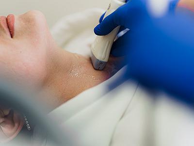
In practice, the ultrasound procedure is fully comfortable: you simply lie on the examination couch while the physician smoothly glides a plastic transducer across the skin. On the screen, it appears as a real-time black-and-white image where dense structures (such as bones) look white, soft tissues appear gray, and fluids look completely black. When color Doppler mode is activated, blue and red flow signals appear on the screen, illustrating blood movement through the vessels.
To prevent gas in the digestive tract from obstructing sound waves and to ensure clear imaging, certain examinations require brief preparation. Preparation depends entirely on the examined area:
Following these simple guidelines ensures the physician can evaluate all anatomical details clearly. If you have questions regarding preparation, our reception staff will provide full instructions during booking.
To ensure high diagnostic accuracy, every examination in our clinic follows a standardized, comfortable protocol:
The entire procedure is conducted calmly and without haste. The doctor explains the key findings during the examination and advises on the appropriate specialist to consult if any abnormalities are detected.
Our specialists regularly carry out the following ultrasound examinations:
Extensive clinical experience allows us to accurately assess the examined structures.
High-quality ultrasound in Warsaw at „Lekarze w Warszawie” combines advanced medical technology with clinical expertise. Our modern diagnostic department is equipped with expert-class ultrasound units capable of detecting subtle structural changes at early disease stages. Consultations are performed by experienced physicians who explain scan findings clearly during the visit.
You no longer need to search for prompt and reliable diagnostic imaging. At „Lekarze w Warszawie”, appointments are scheduled in advance, preventing waiting times and ensuring dedicated attention for every patient. Convenient morning and evening appointment slots are available.
Our reception team will contact you to confirm your visit, explain all preparation guidelines, and assist with scheduling. You can book an appointment via the website form or by phone.
Yes, it is 100% safe. Ultrasound does not expose the body to ionizing radiation, unlike X-rays. Because it relies entirely on sound waves, it can be performed as often as clinically necessary, including during pregnancy and in newborns from the first days of life.
Typically, examining a single area takes 15 to 30 minutes. A comprehensive multi-system examination may take approximately 45 minutes.
No, preparation is required only for examinations of the abdomen, kidneys, and pelvic organs. Imaging of the neck, thyroid, vessels, lymph nodes, and joints requires no preparation.
The procedure is entirely painless. Patients experience only the cool touch of the acoustic gel and the smooth glide of the transducer. Mild tenderness may occur only if the examined organ is acutely inflamed and direct pressure is applied.
Ultrasound enables prompt detection of inflammation, organ enlargement, benign and malignant lesions, gallbladder stones, kidney stones, and vascular disorders such as stenosis, plaques, and thrombosis. Timely imaging facilitates prompt treatment planning before severe complications develop.
An ultrasound should be scheduled in cases of unexplained pain, discomfort, abnormal blood tests, or as part of routine annual checkups. In challenging clinical scenarios where standard imaging is inconclusive, contrast-enhanced ultrasound (CEUS) may be indicated. Microbubble contrast agents enhance vascular visualization, allowing precise differentiation between benign and malignant lesions.
We care for your health with full commitment and professionalism.
Abdominal ultrasound
zł 270.00
Thyroid ultrasound
zł 250.00
Genitourinary ultrasound
zł 250.00
Salivary gland ultrasound
zł 280.00
Ultrasound package: Abdomen + urinary tract
zł 410.00
Thyroid Ultrasound
"2 Ultrasound Scans" Package (excluding heart and vessels)
Genitourinary ultrasound
zł 250.00
"3 Ultrasound Scans" Package (excluding heart and vessels)
zł 600.00
Lung ultrasound
zł 270.00
Soft tissue ultrasound (2 areas)
zł 230.00
Lymph node group ultrasound (1 area)
zł 290.00
Behind the success of our clinic are experienced doctors – experts in their fields who combine knowledge, passion, and an individual approach to the patient.
What our clients say about us
Nina Zastavna

We had an ultrasound done for my five-year-old daughter, and everything went great. The staff's approach was simply wonderful; the child felt safe and wasn't afraid. I definitely recommend it.
Xenia Andrushchenko
I was there for an ultrasound - the doctor was very attentive, she checked everything thoroughly and calmly. I recommend!
Sviatlana Areshchankova
I would like to thank the doctors very much for your work! I liked everything. I highly recommend them.
We have answers to all questions
No, a referral is not required. It is enough to make an appointment for the examination by phone or online.
No. The ultrasound examination is completely painless, safe, and non-invasive — it can be performed repeatedly.
It usually takes from 10 to 30 minutes, depending on the area being examined.
Preparation depends on the type of examination. In case of doubt, our staff will be happy to explain how to best prepare for a specific ultrasound.
Ultrasound results are available immediately after the examination — the doctor discusses them right away during the visit.
We perform, among others, ultrasound of the abdomen, genitourinary system, thyroid, soft tissues, lymph node groups, and others.


.jpg)